Research
The Challenge
Cancer and neurodegenerative diseases represent some of the most significant challenges facing modern society. With the WHO predicting a steady rise in these conditions, the need for advanced diagnostic and therapeutic interventions has never been more urgent.
The ADMIT Mission
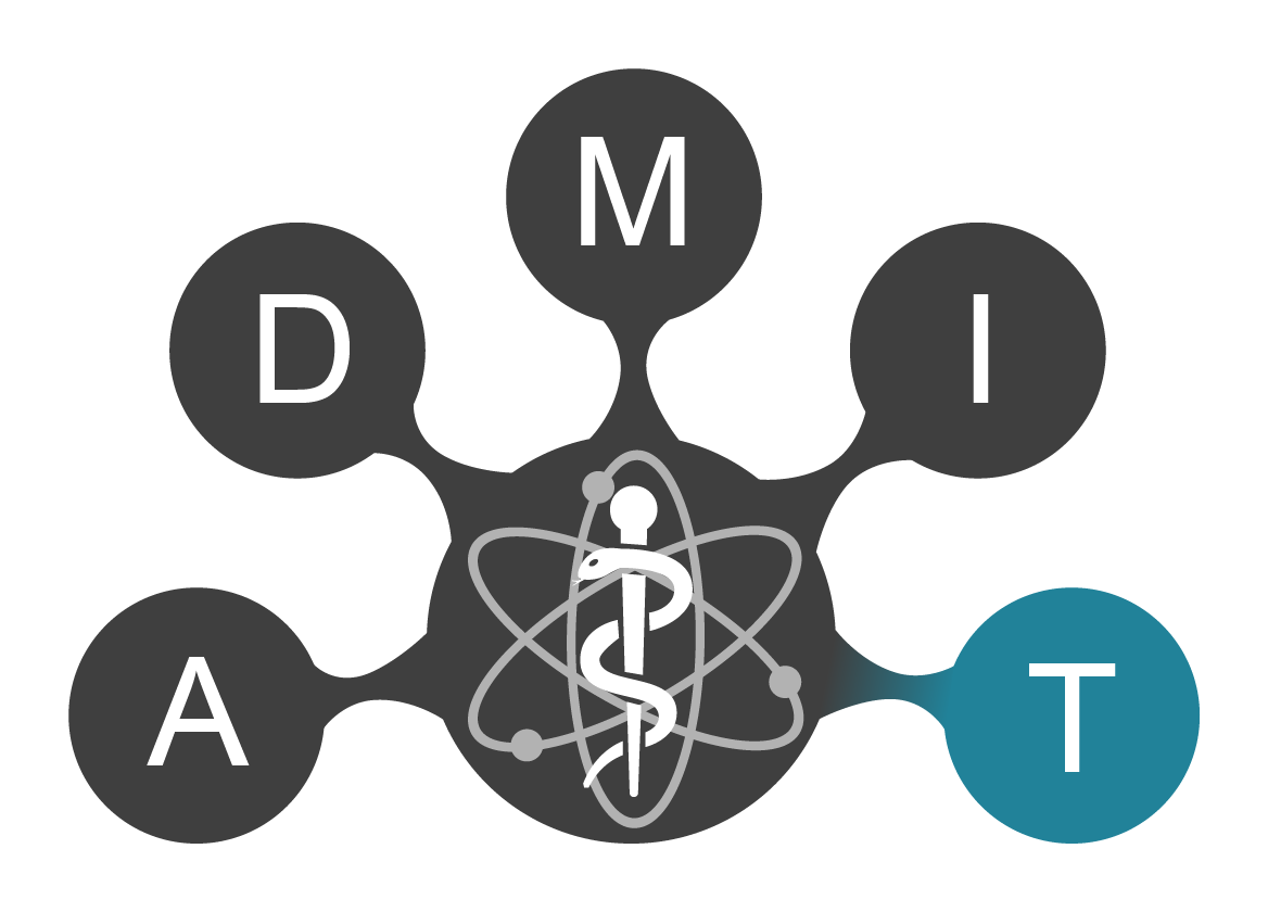
To meet this challenge, the LOEWE priority project ADMIT is at the forefront of medical physics innovation. Our mission is to bridge the gap between theoretical research and clinical application by combining cutting-edge imaging and computer-assisted techniques with advanced therapeutic strategies. By doing so, we aim to enable earlier diagnosis and more successful treatment outcomes for cancer and neurodegenerative disorders.
Our Approach
At the core of ADMIT is the development of novel medical physics methodologies designed to enhance image-guided therapy and unlock new research avenues. We integrate scientifically rigorous approaches into the synergistic fields of data processing, imaging, and clinical therapy—specifically within radiology, oncology, and neurology.
Collaborative Excellence
ADMIT is a powerhouse of interdisciplinary expertise, bringing together researchers from the THM, UMR, and JLU universities. Our strength is amplified through strategic clinical and industrial partnerships, as well as long-standing international collaborations with prestigious institutions including Harvard University, Yale University, and the University of Pennsylvania.
Project Area A | Range Modulator and Neutrons in Particle Therapy with Protons
Pushing the boundaries of FLASH irradiation: We are investigating neutron exposure to ensure the next generation of particle therapy is as safe as it is effective. Challenge FLASH irradiation is a promising method for significantly sparing healthy tissue during high-energy ionising radiation treatment. However, when using protons or carbon ions, fragmentation produces neutrons. Due to their high biological effectiveness, these neutrons pose a non-negligible risk of secondary malignancies. Furthermore, the extent of neutron exposure associated with bolus materials remains unclear for clinical use. Scientific Approach The project combines experimental measurements with Monte Carlo simulations. Local neutron dose distributions and energy spectra are measured using thermoluminescence ...
Project Area D | Monte Carlo Simulations to Describe the Radiation Effect of Carbon Ions and Protons at the Cellular Level
Decoding the impact of radiation: Using advanced Monte Carlo simulations to model biological effects at the cellular level for more precise cancer treatments. The Challenge Particle beams exhibit an elevated relative biological effectiveness (RBE) compared to photon beams. However, calculating RBE is highly complex; current models, such as LEM and MKM, show uncertainties of up to 30%, and they have not yet been clinically established for proton beams. There is a critical need for more precise predictions regarding effects at the cellular level. Scientific Approach The project employs Monte Carlo simulations as an efficient method to characterise the effects of ionising radiation. The focus is on further optimising and experimentally validating complex chemical and biological effects ...
Project Area M | Adaptive Radiotherapy: Deep Learning for the Analysis of Daily Imaging
Smart radiotherapy for personalized care: Leveraging Artificial Intelligence to adapt radiation doses in real-time to a patient's changing anatomy. Challenge Adaptive radiotherapy focuses on a patient's anatomical changes between fractions—such as weight loss or fluctuations in bladder and bowel filling. A key challenge is determining whether a recalculation and re-optimisation of the dose distribution is required to maintain treatment accuracy. Scientific Approach The project aims to develop and validate artificial intelligence neural networks to enable rapid, independent assessment of daily imaging data. This allows for a more efficient evaluation of whether anatomical changes necessitate a change in the radiation plan. Objectives and Impact The objective is to provide medical ...
Project Area I | Spectral Computed Tomography and Low-Dose Image Reconstruction Techniques
Clearer images, lower dose: Revolutionising CT imaging through spectral technology to improve diagnostics while significantly reducing radiation exposure. Challenge The increasing frequency of CT examinations leads to higher radiation exposure across the population. Furthermore, the presence of metallic implants creates significant issues, such as beam starvation and streaks, which can compromise image quality and render images non-evaluable. Scientific Approach Modified imaging techniques combined with spectral CT are being developed to improve diagnostic quality while simultaneously reducing radiation exposure. Key objectives include improving the signal-to-noise ratio for better tissue differentiation, reducing metal-induced artefacts, and investigating ways to reduce the need ...
Project Area T | MR Imaging-Assisted Deep Brain Stimulation Therapy
Bridging the gap between MRI and Deep Brain Stimulation: Developing innovative hardware to make advanced neuroimaging safe and accessible for DBS patients. Challenge A primary challenge in Deep Brain Stimulation (DBS) is ensuring compatibility with Magnetic Resonance Imaging (MRI). The interaction between electromagnetic fields and implanted electrodes can interfere with imaging, limiting the clinical utility of MRI for patients with DBS implants. Scientific Approach The project aims to develop DBS-compatible MRI hardware through innovative engineering. This includes the use of special transmitter coils and the development of add-on systems for parallel transmission at 3 Tesla field strength to reduce electromagnetic interference. The process involves simulation-based development ...